Researchers, including one of Indian-origin, have successfully produced the first 3-D anatomic model of a patient's heart using two common imaging techniques, aiming to enhance diagnosis and surgical planning.
The 3D model printing of patients' hearts has become more common in recent years as part of an emerging, experimental field devoted to enhanced visualisation of individual cardiac structures and characteristics.
But this is the first time the integration of computed tomography and three-dimensional transesophageal echocardiography has successfully been used for printing a hybrid 3D model of a patient's heart.
"Hybrid 3D printing integrates the best aspects of two or more imaging modalities, which can potentially enhance diagnosis, as well as interventional and surgical planning," said Jordan Gosnell, from Helen DeVos Children's Hospital in the United States, and lead author of the study.
"Previous methods of 3D printing utilise only one imaging modality, which may not be as accurate as merging two or more datasets," said Gosnell.
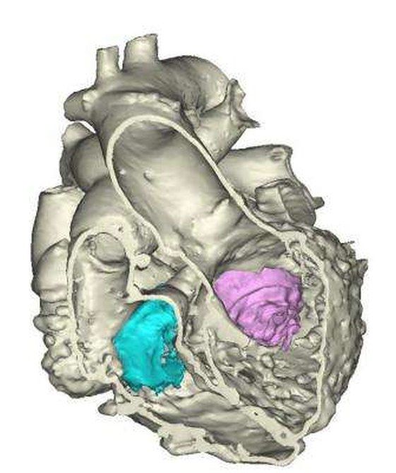
The team used specialised software to register images from the two imaging modalities to selectively integrate datasets to produce an accurate anatomic model of the heart. The result creates more detailed and anatomically accurate 3D renderings and printed models, which may enable physicians to better diagnose and treat heart disease.
CT and magnetic resonance imaging are established imaging tools for producing 3D printable models. The 3DTEE recently was reported by Joseph Vettukattil, and his Helen DeVos Children's Hospital colleagues to be a feasible imaging technique to generate 3D printing in congenital heart disease.
According to Vettukattil, senior author of the study, and his colleagues, each imaging tool has different strengths, which can improve and enhance 3D printing.
3DTEE provides the best visualisation of valve anatomy, he said.
"This is a huge leap for individualised medicine in cardiology and congenital heart disease," said Vettukattil.
"The technology could be beneficial to cardiologists and surgeons. The model will promote better diagnostic capability and improved interventional and surgical planning, which will help determine whether a condition can be treated via transcatheter route or if it requires surgery," Vettukattil added.
Image courtesy: Materialise